


The eye has been called the most complex organ in the body. It's amazing that something so small can have so many working parts. But when you consider how difficult the task of providing vision really is, perhaps it's no wonder after all.
Click on the words below for their definitions.
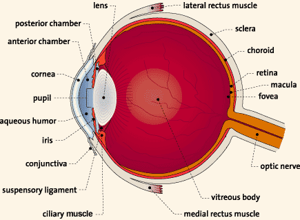
The eye is like a camera. It lets light in through the cornea, which is like a camera's opening. The amount of light allowed in is controlled by the pupil, which opens and closes a bit like a shutter. The light focuses on the retina, which sends the image to the brain, acting as film would in order to record the light (the photo itself).
Other eye structures support the main activity of sight. Some carry fluids - tears and blood - to lubricate or nourish the eye. Others are muscles that allow the eye to move. Some protect the eye from injury - lids and the epithelium of the cornea. And some are messengers, sending sensory information to the brain - pain-sensing nerves in the cornea and the optic nerve behind the retina.